Pause
Play
This is only the beginning...
A lot happens between giving a sample and getting results. Scroll down to learn more about how the laboratory is involved with your health.
A lot happens between giving a sample and getting results. Scroll down to learn more about how the laboratory is involved with your health.
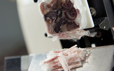
This is a Histotechnological stain applied on skin tissue which includes a hair follicle. The actual name of this stain is Hematoxylin and Eosin (better known as H&E). It is a routine stain applied to tissues received in the Histopathology lab. ALL tissues removed from the patients (surgical, biopsy, autopsy) are sent to the Histopathology lab.
Once received in the lab, medical laboratory professionals put the samples through a series of processes, including staining. After applying stains, the tissue components will pick up the dye in the staining solution making them visible under the microscope. The medical laboratory professional ensures the samples are processed correctly before handing them to a pathologist for a diagnosis. This is the only way the tissues from the patient can be diagnosed; through the visualization of the tissue architecture by the pathologist.
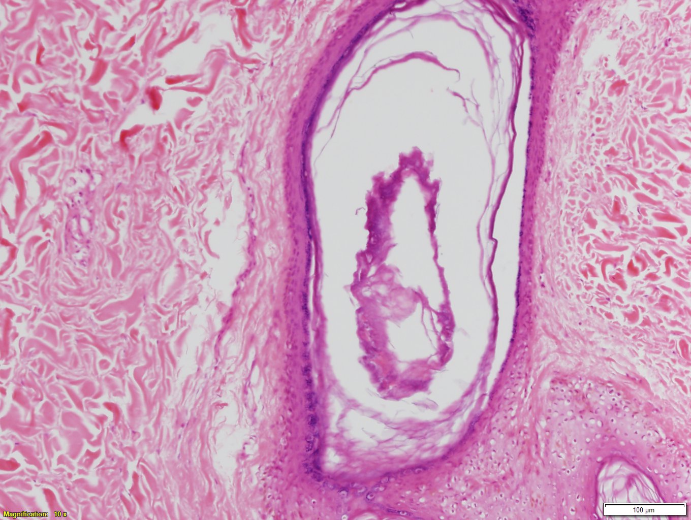
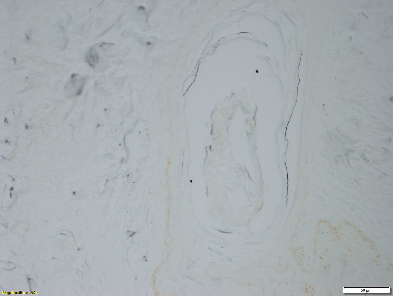
courtesy of The Michener Institute Histology Faculty
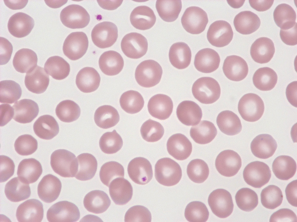
Blood can tell many stories. Blood tests are often ordered through a routine health exam or when there are symptoms that require further investigation.
A medical laboratory professional will view the prepared blood sample under a microscope. How the blood cells appear is important information used in diagnosis.
Credit: Sysmex Corporation
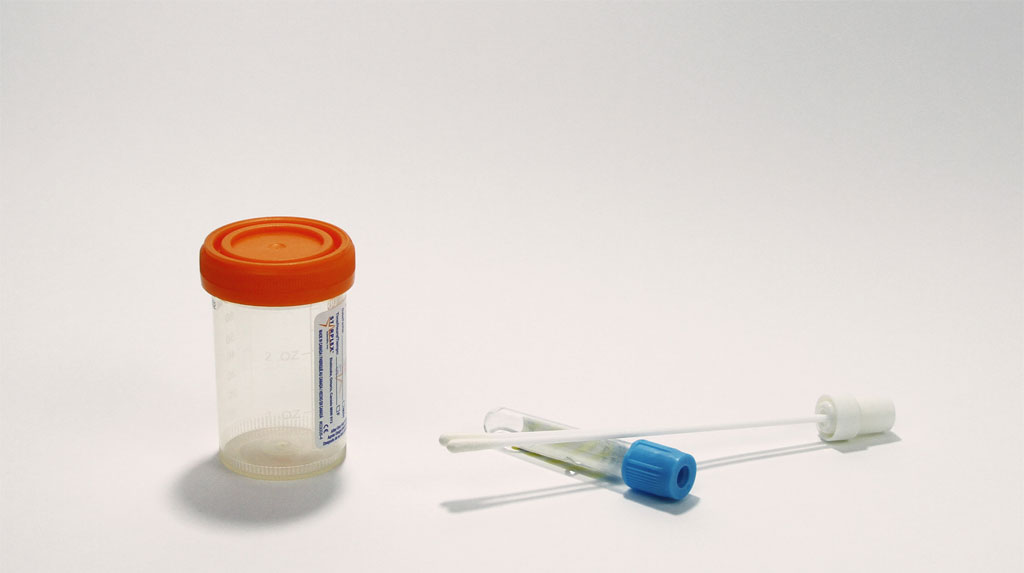
Bacteria grow in your body causing an infection, making you feel ill or in pain. A very common bacterial infection is strep throat, caused by Streptococcus A.
Your doctor or nurse will take a throat swab to get a sample of the bacteria cells in your throat. Then the sample is sent to the lab to grow the bacteria to determine the type, in order to know the treatment.
A medical laboratory professional will take a very small part of the sample and touch it to a petri dish that has been treated to enable bacteria growth. They will then use a small, sterile instrument to spread the cells around the dish, this is called streaking. It helps to separate as many cells as possible.
Once the bacteria begins to grow it may look like this in the petri dish.
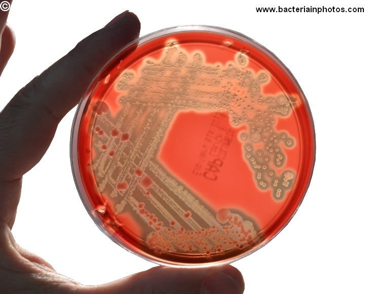
courtesy of bacteriainphotos.com
The medical laboratory professional will then take a small part of the growing bacteria and process it, including staining (or adding colour) in order to view the cells under a microscope. This helps determine what strain or type of bacteria is present and which antibiotic will treat it.
Depending on the results of blood work and ultrasounds, a primary physician may recommend a liver biopsy. This might be for patients with a history of alcohol abuse or those who have had a hepatitis infection at one point. They will likely have their liver function monitored by a specialist on a regular basis so that cirrhosis will not ensue or progress further.
In this sample we see adipose tissue (fat) infiltrating the normal architecture of the liver specimen, seen as the large white cells. The presence of fat amongst the normal liver architecture is a tell-tale sign that you are visualizing a progressed disease. A fatty liver can be the result of several diseases or disorders including alcohol abuse, obesity and hepatitis.
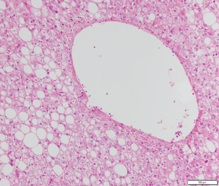
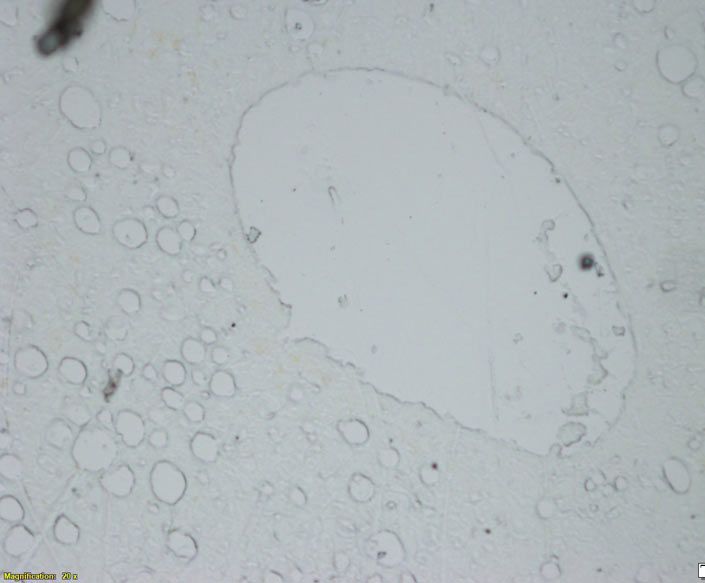
courtesy of The Michener Institute Histology Faculty
A medical laboratory professional would process the biopsy sample in order for a pathologist to make a visual diagnosis. You can see the importance of staining a sample, in order to see the various structures.
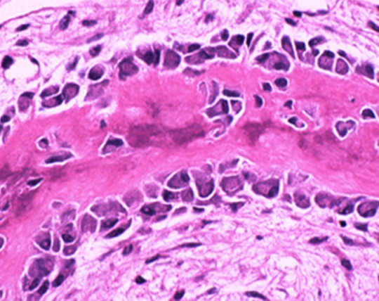
Osteoblasts are one of the cell types that form bone. They are seen here in a bone biopsy. A biopsy might be performed to check for infection, cancer or other bone diseases. This image is of normal bone that has been decalcified (removal of the hard calcium), processed and stained (coloured) by laboratory professionals and magnified under a microscope. It will be given to a pathologist for examine and possible diagnosis.
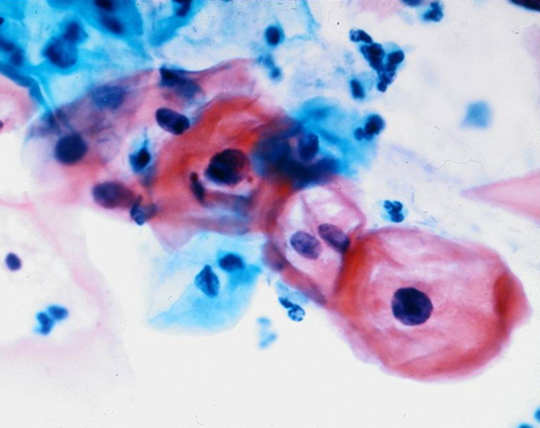
This image shows squamous epithelial cells infected with Human Papilloma Virus (HPV). These cells are from a Pap test (Papanicolaou test). This routine test involves taking a sample from a woman's cervix to screen for cervical cancer. The sample is processed, stained (coloured) and examined by laboratory professionals.
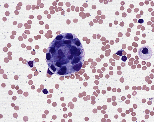
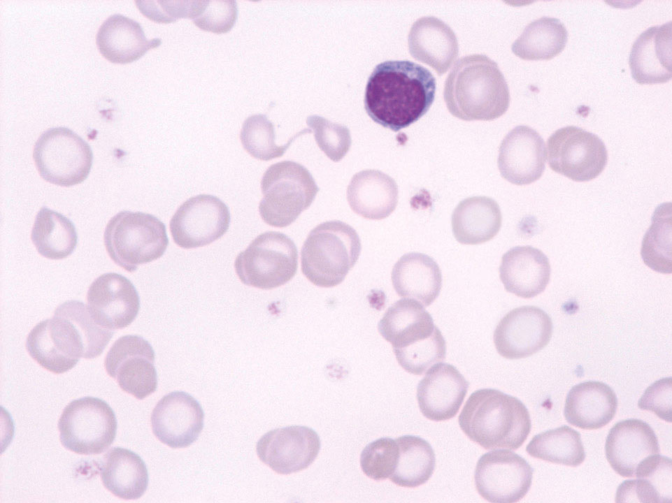
Pleural fluid is found in the membranes surrounding your lungs. It's normal to have a small amount of this fluid as it helps us breathe normally. With certain diseases such as lung cancer, too much fluid is produced. This is a picture of pleural fluid from someone with metastatic lung cancer. The large purple cell in the middle is a cancerous cell. Laboratory professionals prepare, stain and examine this type of fluid and identify the cells found in it. If a cell looks suspicious it is referred to a pathologist for final diagnosis.
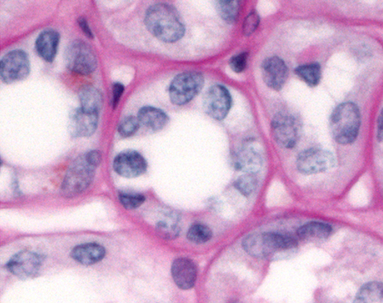
Renal tubules are the small tubes found in your kidneys that contain fluid filtered by the kidneys. As fluid travels through these tubules it is further filtered. Anything your body requires such as water, electrolytes, glucose and amino acids are kept and anything that is not needed is excreted as urine. This image is of a normal renal tubule has been processed and stained (coloured) by laboratory professionals and magnified under a microscope. A renal tubule may appear abnormal in diseases such as systemic lupus erythematosus or with kidney failure.
Sometimes diagnosing the cause of illness can require several tests. The combination of results from x-ray, ultrasound and blood testing might only provide part of the story. Your doctor may need a closer look at what's happening under the skin surface and that will lead to a tissue biopsy.
A sample of tissue is removed from the body (biopsy) and is prepared for visual analysis under the microscope by a pathologist. A medical laboratory professional in the histology lab, performs several steps to ensure the sample is prepared correctly for the most accurate results.
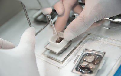
Step 1: The tissue is processed to dehydrate it and then embedded into a wax mold.
Step 2: The wax-embedded tissue is sliced very finely to reveal deeper layers

Step 3: The thin layers of wax-embedded tissue is mounted on a microscope slide and stained to enhance details

Step 4: The slide is viewed under the microscope where normal and abnormal cells can be viewed.
The demand for laboratory tests is always increasing. The volume of some tests have risen exponentially. In the case of vitamin D tests, the number of tests ordered have increased dramatically, driven by the interest of both doctors and their patients in the purported health benefits of the vitamin.
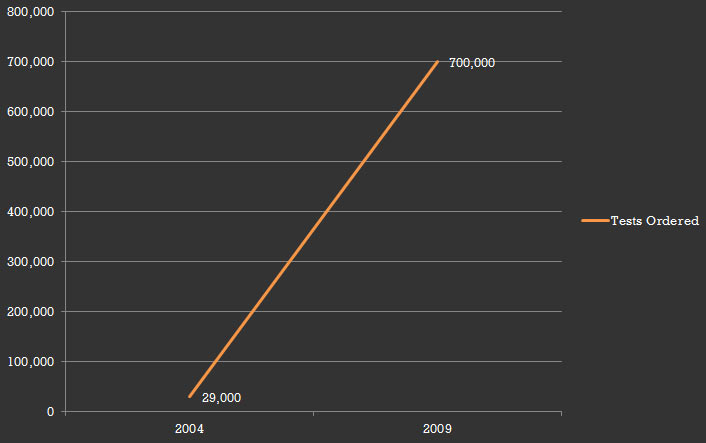
The number ordered by doctors rose to about 700,000 in 2009 from 29,000 in 2004.1
Laboratory tests are required for an accurate diagnosis of the specific bacteria causing a sexually transmitted infection (STI). This information is vital to providing effective treatment, as each bacteria would respond to a different antibiotic.

In 2010, there were:
94,690 reported cases of chlamydia, 11,397 reported cases of gonorrhea, and 1,757 reported cases of infectious syphilis.2
The diagnosis of cancer in any part of the body can be a multi-step process, which can include x-ray, ultrasound, blood, genetic and chemical testing. The laboratory plays a vital role in this process. The testing done in the laboratory are key to assisting in early detection and treatment.
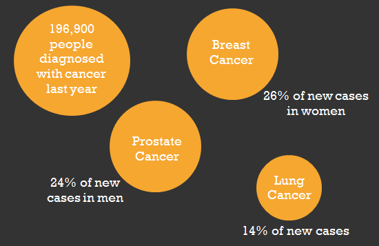
196,900 people diagnosed with cancer last year3
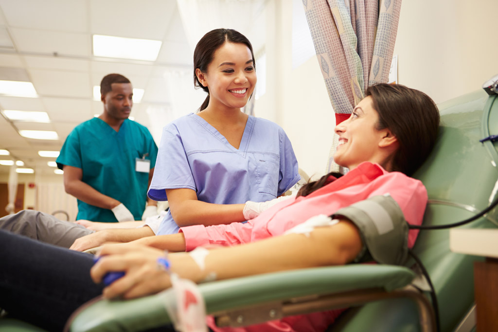
In one year 850,218 blood donations were received.4
Donated blood is used for many life-saving treatments. When blood and blood products are donated, they are carefully screened in the lab to ensure they are safe to use. When patients require blood, the medical laboratory professionals in transfusion medicine will check for the compatibility of blood products being used. Every product is tested in the lab for safety and compatibility.

Amniocentesis (also referred to as amniotic fluid test or AFT) is a test in which fetal DNA is examined for genetic abnormalities. 10,000 Canadian women undergo amniocentesis each year10

Almost every hour, more than 20 people are newly diagnosed with diabetes. Each diagnosis is made through a laboratory blood test called Hemoglobin A1c.
A medical laboratory professional processes a blood sample to determine the level of glucose (sugar) that is present in the blood. They can tell this by the level of glycated hemoglobin (or A1c) that is present, because A1c is present when glucose attaches to hemoglobin.
Once a diagnosis is made, diabetes can be monitored and managed with the help of continual laboratory tests.8

Early detection is an important factor in beating cancer.
People diagnosed with cancer today have a better five-year relative survival than they did just over a decade ago. Between 1992 to 1994 and 2006 to 2008, the five-year relative survival for all cancers combined increased by 7.3 percentage points from 55.5% to 62.8%.

Leukemia estimates for 2015: 5
The rates of incidences and death rates of cervical cancer has significantly declined over several years due to regular screening with Pap tests. This laboratory test has been vital to early detection and treatment.6
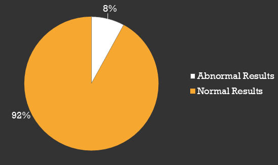
Approximately 4,000,000 Pap tests are done every year in Canada. Around 1,400 women are diagnosed with cancer from these abnormal results.7
When your primary health care provider is looking for answers, they may turn to laboratory tests. Highly trained medical laboratory professionals sort, analyze and run tests on your samples to provide accurate results that are vital to your health care.
Testing done in the medical laboratory is an important part of your health. The results from laboratory testing can lead to the diagnosis and treatment of illness or disease in every part of your body.
Discover how medical laboratory professionals are involved in your health. Click on the points to learn about some of the testing done on that body part.

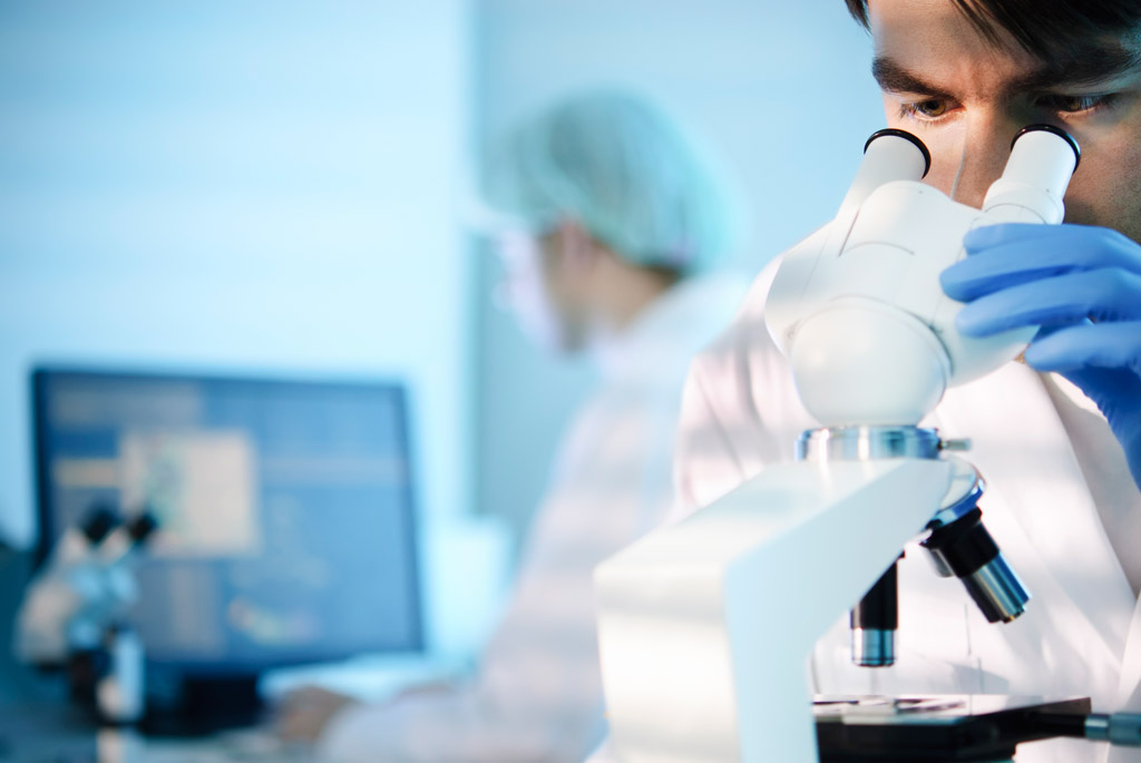
Medical laboratory professionals work around the clock to provide accurate and timely results vital to medical decisions about your health. These health care professionals use a variety of instruments and equipment to collect, prepare and analyze tissue and fluid samples.
Hover your cursor over each professional below to learn more about them.

Medical Laboratory Technologists (MLTs) use a variety of complex instruments to analyze tissue samples, blood and other body fluids as a part of the diagnostic procedure. MLTs provide the results of these sophisticated tests to physicians, allowing them to make accurate diagnosis and if needed, appropriate treatment.
Medical Laboratory Assistants (MLAs) work under the supervision of a Medical Laboratory Technologist (MLT), performing the practical components of sample analysis. MLAs sort, prepare and sometimes process samples that will be tested and analyzed by a MLT. MLAs often collect samples, such as blood, and are the laboratory professional that likely interacts directly with patients.
Cytotechnologists are health professionals that analyze cellular changes that can determine the presence of specific diseases. Mostly through the use of slides under a microscope, cytotechnologists are able to detect pre-cancerous cells, different cancers and other cellular based infections. An abnormal finding would be sent to a pathologist for a final diagnosis.

Genetic Technologists use a variety of instruments to analyze and diagnose changes or abnormalities in chromosomes and DNA, which are unique to every individual. A genetic technologist's analysis of these cells can lead to a diagnosis of genetic diseases.
As a patient, the care you receive is supported by medical laboratory professionals. You may never see them, but their impact on patient care is bigger than you may think. They work around the clock to provide accurate and timely results vital to your health – giving you the answers you need, when you need them most.
There are over 440 million lab tests performed on a yearly basis in Canada. Discover how lab tests make a difference by clicking on each discipline below.


Clinical Chemistry

Clinical Microbiology
Hematology
Diagnostic Cytology

Clinical Genetics
Transfusion Science
Histology
Clinical Chemistry
Medical laboratory professionals are proud of the work they do and how their job contributes to patient care. This is what few medical laboratory professionals had to say about their role in health care.
MLTs are a catalyst for patients being diagnosed and treated so they can feel good again.


Jennifer O'Neill
I am proud to represent a profession that plays an integral part in patient care and diagnosis!


Krista Urchenko
The ability to perform a procedure such as phlebotomy while making someone smile is what I love most about my job. I do my best to create an encouraging atmosphere that allows patients to feel comfortable.

Samantha Tanner
The thing I love most about being an MLT, is the variety of disciplines to work in. There is always something new to learn and discover.

Evelyn Aboagye
My contribution to patient health and well-being really makes a difference.

Mary Emes
I am happy to be able to help my community members in their times of need when they are sick.

Kim Mathison


Help us raise awareness about the important work done by medical laboratory professionals in Canada. Please share our message.
#LabWeek